

THP-1 Human NFκB-Luc Reporter Cell
Cat. No: RQPB0013
Size: 1 vial of frozen cells (>1E6 per vial in 1 mL)
Unit Price: Contact For Pricing
Product Info
Description
Assay Data
Cell Culture
| Cat. No | RQPB0013 |
| Product Name | THP-1 Human NFκB-Luc Reporter Cell |
| Product Type | Reporter Cell |
| Culture Properties | Suspension |
| Stability | 32passages (in-house test, that not means the cell line will be instable beyond the passages we tested.) |
| Mycoplasma Status | Negative |
| Culture Medium | RPMI-1640+10%FBS+1μg/ml puromycin+0.05 mM 2-mercaptoethanol |
| Freeze Medium | 90% FBS+10% DMSO |
| Storage Conditions | Liquid nitrogen immediately upon delivery |
For research use only. Not intended for human or animal clinical trials, therapeutic or diagnostic use.
Nuclear Factor kappa-light-chain-enhancer of activated B cells (NF-κB) is a family of transcription factor protein complexes that controls DNA transcription, cytokine production, and cell survival. NF-κB is present in almost all animal cell types and participates in cellular responses to stimuli such as stress, cytokines, free radicals, heavy metals, ultraviolet irradiation, oxidized LDL, and bacterial or viral antigens.
NF-κB activation involves two major signaling pathways: the canonical pathway and the non-canonical (or alternative) pathway. Although their signaling mechanisms differ, both are crucial for regulating immune and inflammatory responses. The primary mechanism of canonical NF-κB activation involves the inducible degradation of IκBα, triggered by site-specific phosphorylation mediated by the multi-subunit IκB kinase (IKK) complex. In contrast to the canonical pathway, the non-canonical NF-κB pathway responds selectively to a specific subset of stimuli—specifically, ligands for certain members of the TNFR superfamily, such as LTβR, BAFFR, CD40, and RANK.
NF-κB activation is initiated when molecules such as TNFα bind to TNF receptors (of which there are various types). Once a TNF receptor is activated, a complex signal transduction cascade ensues; the IκB kinase (IKK) is ultimately triggered, leading to the phosphorylation of IκB, which in turn results in the ubiquitination and degradation of IκB. Upon the degradation of IκB, the remaining NF-κB dimers (e.g., p65/p50 or p52/p50 subunits) translocate into the cell nucleus, where they bind to consensus DNA sequences within target genes.
NFκB-Luc THP-1 reporter cells are THP-1 cells engineered to express a luciferase (Luc) reporter gene under the transcriptional control of NF-κB. The principle by which TNFα binding to its receptor activates NF-κB is illustrated in the figure below.

Figure 1. Schematic Diagram of the NFκB-Luc THP-1 Principle
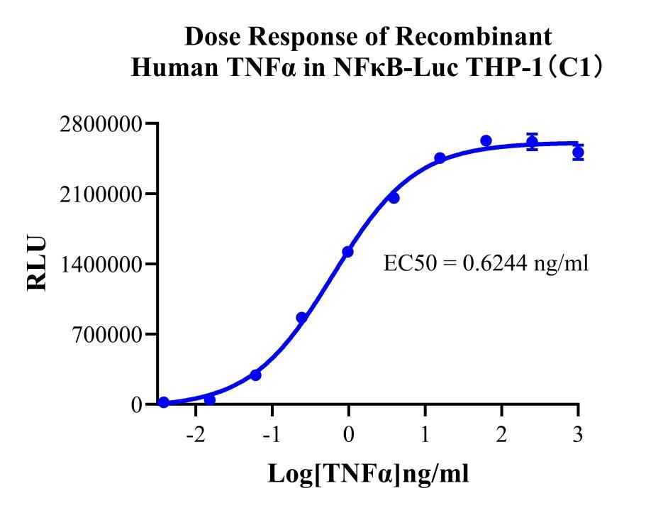
Figure 2. Dose Response of Recombinant Human TNFα in NFκB-Luc THP-1(C1).
Cell Passage Procedures
1.This cell line grows in suspension.
2.Upon receipt, cells should be thawed immediately or stored in liquid nitrogen until use.
3.Before thawing, pre-warm the water bath and culture medium to 37 °C, and prepare a small amount of dry ice.
4.Remove the cryovial from storage and transport it to the cell culture laboratory on dry ice.
5.Rapidly thaw the cells in a 37 °C water bath. Once the cells are completely thawed, spray the cryovial with 70% ethanol for disinfection and transfer it to a biosafety cabinet.
6.Add 10 mL of pre-warmed culture medium into a 15 mL centrifuge tube. Transfer the contents of the cryovial into the tube and centrifuge at 1000 rpm for 5 minutes.
7.Carefully discard the supernatant. Resuspend the cell pellet in 5 mL of pre-warmed culture medium by gentle pipetting. Immediately perform cell counting and adjust the cell density to 3–6 × 10⁵ cells/mL based on the counting results, then transfer the cells into a culture flask.
8.Count the cells every 1–2 days. When the cell density exceeds 1 × 10⁶ cells/mL, passage the cells promptly or add fresh culture medium. Maintain the cell density between 2 × 10⁵ and 1 × 10⁶ cells/mL.
Suspension Cell Cryopreservation Procedure:
1.Collect 8 × 10⁶ cells, centrifuge, and discard the supernatant.
2.Add 1 mL of cell freezing medium (90% FBS + 10% DMSO) and gently pipette to mix thoroughly. Transfer the suspension into a cryovial.
3.Immediately place the cryovial into a controlled-rate freezing container (Nalgene 5100-0001), fill with isopropanol up to the indicated level, and store at −80 °C.
4.After 24 hours, transfer the cryovial to liquid nitrogen for long-term storage.
Related products
HEK293 Human NK1R CRE-Luc Cell Line
Raji-Luc-GFP
Jurkat E6.1-Luc
THP-1-GFP
THP-1-Luc
Raji-GFP
Raji-Luc
Jurkat E6.1-GFP
HEK293 Human GAL4-Luc Reporter Cell
HEK293 Human CRE-Luc Reporter Cell
We Are Pleased to Announce: Global Commercial Licensing Rights for Jurkat E6.1, CHO-K1, and HEK293 Cell Lines Officially Secured.
Explore