

Jurkat E6.1 Human KIR3DL3 Effector Reporter Cell
Cat. No: RQP74193
Size: 1 vial of frozen cells (>1E6 per vial in 1 mL)
Unit Price: Contact For Pricing
Product Info
Description
Biological Information
Assay
Cell Culture
| Cat. No | RQP74193 |
| Product Name | Jurkat E6.1 Human KIR3DL3 Effector Reporter Cell |
| Product Type | Reporter Cell |
| Culture Properties | suspension |
| Stability | 32passages (in-house test, that not means the cell line will be instable beyond the passages we tested.) |
| Mycoplasma Status | Negative |
| Culture Medium | RPMI-1640+10%FBS+1μg/ml puromycin+800μg/ml Hygromycin B |
| Freeze Medium | 90% FBS+10% DMSO |
| Storage Conditions | Liquid nitrogen immediately upon delivery |
| Application | Functional(Report Gene) Assay |
For research use only. Not intended for human or animal clinical trials, therapeutic or diagnostic use.
KIR3DL3 (Killer-cell Immunoglobulin-like Receptor 3DL3) is a member of the Killer-cell Immunoglobulin-like Receptor (KIR) family and belongs to the inhibitory receptor subclass. Members of the KIR family facilitate both innate and adaptive immune responses through their expression on NK cells and T cells. KIR3DL3 comprises three extracellular immunoglobulin-like domains, a transmembrane region, and an intracellular segment containing an Immunoreceptor Tyrosine-based Inhibitory Motif (ITIM). KIR3DL3 is primarily expressed on Natural Killer (NK) cells and certain T-cell subsets (such as CD8+ T cells); it principally regulates NK cell activity and plays a balancing role in maintaining immune tolerance as well as in anti-tumor and anti-infection immunity.
HHLA2 (also known as B7H5, B7H7, or B7y) is a member of the B7 family. It is a type I transmembrane protein consisting of an extracellular region featuring tandem IgV1-IgC-IgV2 domains, a transmembrane region, and a cytoplasmic tail. HHLA2 is expressed at low levels in tissues such as the placenta and intestinal epithelial cells, but is highly expressed in various solid tumors (including lung cancer, breast cancer, colorectal cancer, and renal cancer) and within inflammatory microenvironments. HHLA2 possesses dual immunomodulatory functions: it binds to the inhibitory receptor KIR3DL3 (on NK cells) and an unidentified T-cell receptor to transmit inhibitory signals, thereby attenuating anti-tumor immune responses; conversely, it activates the PI3K-AKT pathway via the receptor TMIGD2, thereby promoting T-cell survival and memory formation.
KIR3DL3 exhibits weak binding affinity for classical MHC Class I molecules, such as HLA-A and HLA-B. Upon binding to HLA, the intracellular ITIM motifs of KIR3DL3 are phosphorylated by Src-family kinases (such as Lck). These phosphorylated ITIMs recruit SH2-domain-containing tyrosine phosphatases (SHP-1/SHP-2), which subsequently dephosphorylate activating signaling molecules (such as Syk and Vav1), thereby blocking NK cell degranulation and cytokine (IFN-γ) secretion.
The Jurkat E6.1 Human KIR3DL3 Effector Reporter Cell Model—effectively simulates the signal transduction process of KIR3DL3 *in vivo*. The underlying principle is illustrated in the figure below.
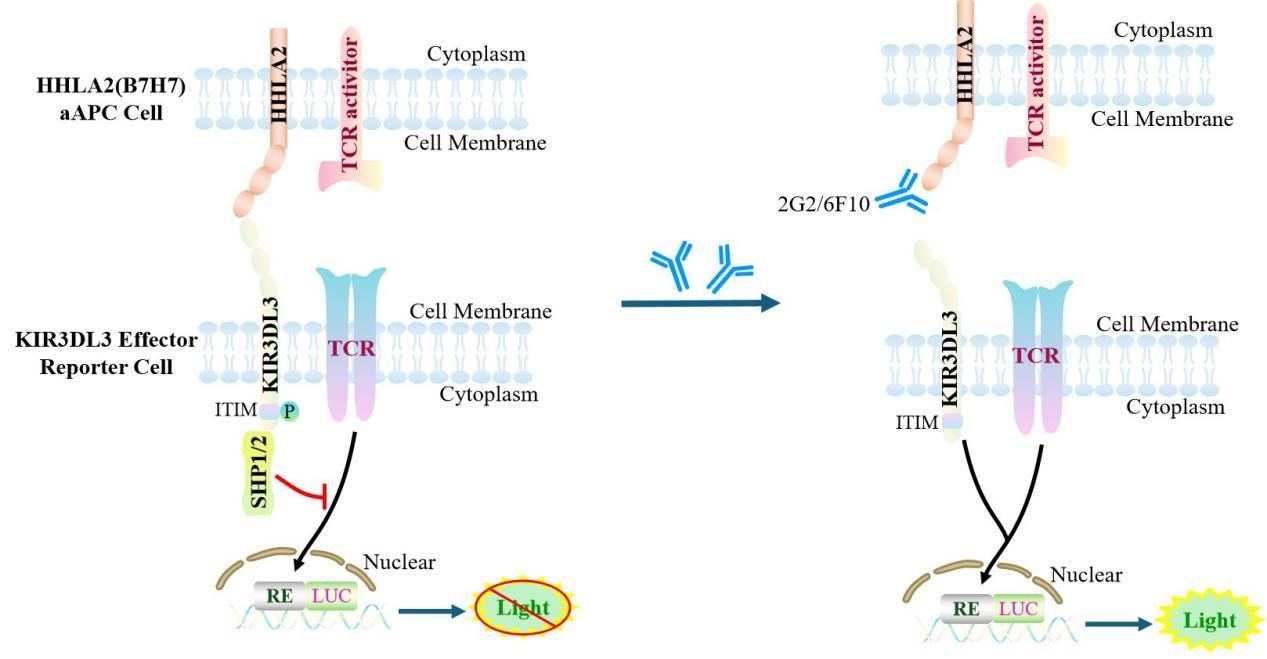
Figure 1. Schematic Diagram of the Jurkat E6.1 Human KIR3DL3 Effector Reporter Cell Model
| Classification | Co-Inhibitory |
| Family | immunoglobulin superfamily |
| Gene Name | KIR3DL3 |
| Gene Aliases | KIRC1;KIR3DL7;KIR44; |
| Gene ID | 115653 |
| Accession Number | NM_153443.5 |
| UniProt Number | Q8N743 |
| Protein Name | Killer cell immunoglobulin-like receptor 3DL3 |
| Protein Aliases | CD158 antigen-like family member Z;Killer cell inhibitory receptor 1 |
| Target Species | Human |
| Host cell | Jurkat E6.1 |
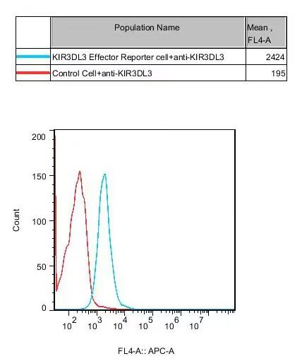
Figure 2. Recombinant KIR3DL3 Effector Reporter Cell stably expressing KIR3DL3.
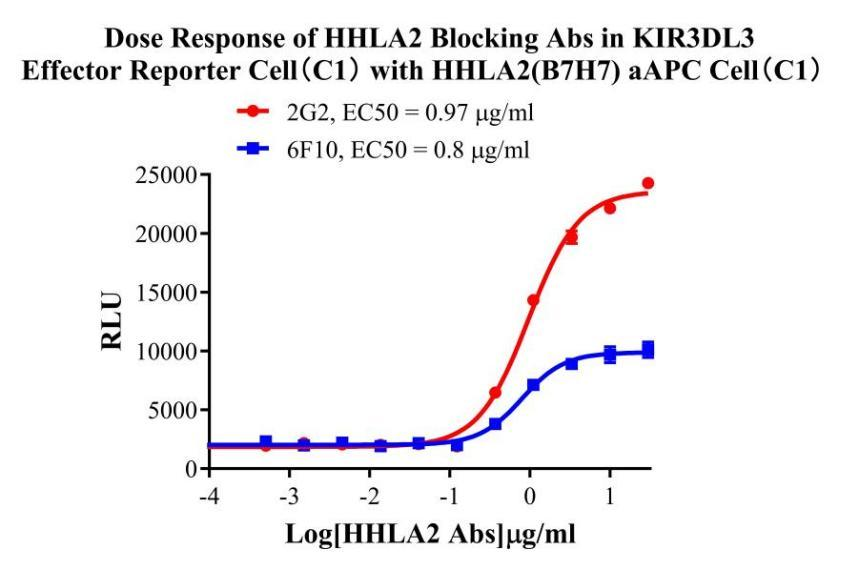
Figure 3. Dose Response of HHLA2 Blocking Abs in KIR3DL3 Effector Reporter Cell (C1) with HHLA2(B7H7) aAPC Cell(C1).
Cell Passage Procedures
1.This cell line grows in suspension.
2.Upon receipt, cells should be thawed immediately or stored in liquid nitrogen until use.
3.Before thawing, pre-warm the water bath and culture medium to 37 °C, and prepare a small amount of dry ice.
4.Remove the cryovial from storage and transport it to the cell culture laboratory on dry ice.
5.Rapidly thaw the cells in a 37 °C water bath. Once the cells are completely thawed, spray the cryovial with 70% ethanol for disinfection and transfer it to a biosafety cabinet.
6.Add 10 mL of pre-warmed culture medium into a 15 mL centrifuge tube. Transfer the contents of the cryovial into the tube and centrifuge at 1000 rpm for 5 minutes.
7.Carefully discard the supernatant. Resuspend the cell pellet in 5 mL of pre-warmed culture medium by gentle pipetting. Immediately perform cell counting and adjust the cell density to 3–6 × 10⁵ cells/mL based on the counting results, then transfer the cells into a culture flask.
8.Count the cells every 1–2 days. When the cell density exceeds 1 × 10⁶ cells/mL, passage the cells promptly or add fresh culture medium. Maintain the cell density between 2 × 10⁵ and 1 × 10⁶ cells/mL.
Suspension Cell Cryopreservation Procedure:
1.Collect 8 × 10⁶ cells, centrifuge, and discard the supernatant.
2.Add 1 mL of cell freezing medium (90% FBS + 10% DMSO) and gently pipette to mix thoroughly. Transfer the suspension into a cryovial.
3.Immediately place the cryovial into a controlled-rate freezing container (Nalgene 5100-0001), fill with isopropanol up to the indicated level, and store at −80 °C.
4.After 24 hours, transfer the cryovial to liquid nitrogen for long-term storage.
Related products
CHO-K1 Human CCR4 Cell Line
HEK293 Human NK1R CRE-Luc Cell Line
Raji-Luc-GFP
Jurkat E6.1-Luc
THP-1-GFP
THP-1-Luc
Raji-GFP
Raji-Luc
Jurkat E6.1-GFP
HEK293 Human GAL4-Luc Cell
We Are Pleased to Announce: Global Commercial Licensing Rights for Jurkat E6.1, CHO-K1, and HEK293 Cell Lines Officially Secured.
Explore