

Jurkat E6.1 Human GARP/Latent TGF-β1 Target Cell
Cat. No: RQP74395
Size: 1 vial of frozen cells (>1E6 per vial in 1 mL)
Unit Price: Contact For Pricing
Product Info
Description
Biological Information
Assay Data
Cell Culture
| Cat. No | RQP74395 |
| Product Name | Jurkat E6.1 Human GARP/Latent TGF-β1 Target Cell |
| Product Type | Reporter Cell |
| Culture Properties | suspension |
| Stability | 32passages (in-house test, that not means the cell line will be instable beyond the passages we tested.) |
| Mycoplasma Status | Negative |
| Culture Medium | RPMI-1640+10%FBS+1 μg/ml Puromycin+10 μg/ml Blasticidin |
| Freeze Medium | 90% FBS+10% DMSO |
| Storage Conditions | Liquid nitrogen immediately upon delivery |
| Application | Functional(Report Gene) Assay |
For research use only. Not intended for human or animal clinical trials, therapeutic or diagnostic use.
GARP (Glycoprotein A Repetitions Predominant) and Latent TGF-β1 (the latent form of Transforming Growth Factor-β1) are closely linked key molecules involved in immunomodulation; together, they play a critical role in maintaining immune homeostasis and influencing the pathogenesis and progression of diseases. The GARP gene is located on human chromosome 9q34.3 and encodes a transmembrane protein belonging to the Leucine-Rich Repeat (LRR) family. Its structure comprises multiple LRR domains, which confer upon it the ability to bind to specific ligands. Latent TGF-β1 is the precursor form of TGF-β1; it is generated through the processing of the TGF-β1 precursor protein into a complex comprising mature TGF-β1, the Latency-Associated Peptide (LAP), and potentially the GARP protein. This complex exists in an inactive state and requires activation before it can exert its biological effects.
The primary function of GARP is to act as a cell- surface receptor that binds and presents Latent TGF-β1. In regulatory T cells (Tregs), GARP is highly expressed; it anchors Latent TGF-β1 to the cell surface, thereby forming a GARP-Latent TGF-β1 complex. When Tregs receive signaling stimuli—such as those mediated by integrins—Latent TGF-β1 can be activated, leading to the release of mature TGF-β1. This release subsequently inhibits the activation of surrounding immune cells, thereby maintaining immune tolerance and preventing autoimmune responses.
Serving as the target cells for the Integrin αvβ6 Effector Reporter Cell system, the GARP/Latent TGF-β1 Target Cells constitute a pharmacological target model that accurately simulates the in vivo signal transduction processes involving Integrins and the GARP/Latent TGF-β1 axis. The underlying principle is illustrated in the figure below.
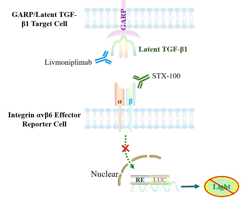
Figure 1. Schematic Diagram of the Jurkat E6.1 Human GARP/Latent TGF-β1 Target Cell Model
| Classification | Cytokine&Growth Factor |
| Family | LRRC32/LRRC33 family |
| Gene Name | LRRC32 |
| Gene Aliases | D11S833E;GARP |
| Gene ID | 2615 |
| Accession Number | NM_001128922.2 |
| UniProt Number | Q14392 |
| Protein Name | Transforming growth factor beta activator LRRC32 |
| Protein Aliases | Garpin;GARP;Leucine-rich repeat-containing protein 32 |
| Family-2 | Transforming growth factor beta family |
| Gene Name-2 | TGFB1 |
| Gene Aliases-2 | TGFB;DPD1 |
| Gene ID-2 | 7040 |
| Accession Number-2 | NM_000660.7 |
| UniProt Number-2 | P01137 |
| Protein Name-2 | Transforming growth factor beta-1 proprotein |
| Protein Aliases-2 | LAP;TGF-beta-1 |
| Target Species | Human |
| Host cell | Jurkat E6.1 |
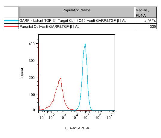
Figure 2. Recombinant GARP/Latent TGF-β1 Target Cell stably expressing GARP&Latent TGF-β1.
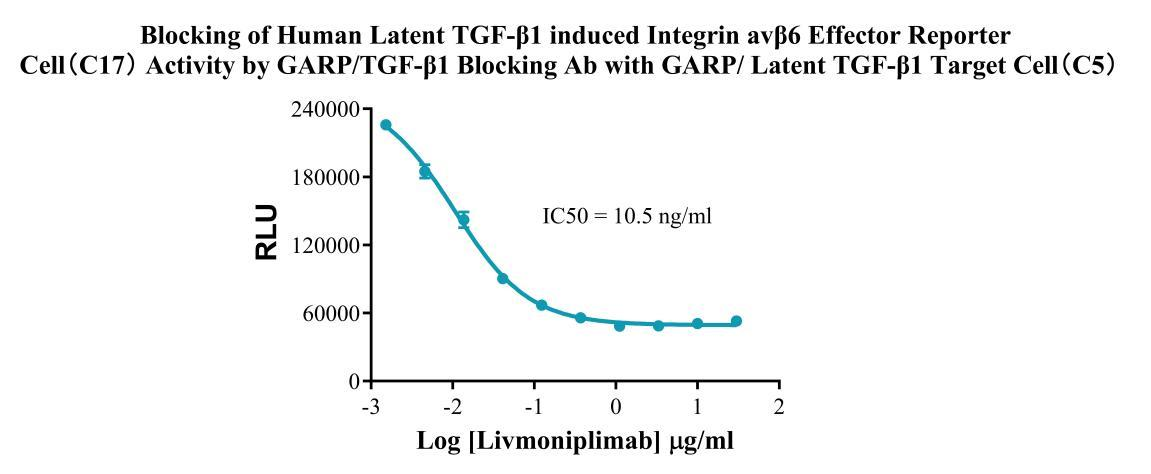
Figure 3. Blocking of Human Latent TGF-β1 induced Integrin avβ6 Effector Reporter Cell(C17) Activity by GARP/TGF-β1 Blocking Ab with GARP/Latent TGF-β1 Target Cell(C5).
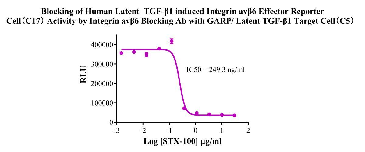
Figure 4.Blocking of Human Latent TGF-β1 induced Integrin avβ6 Effector Reporter Cell(C17) Activity by Integrin avβ6 Blocking Ab with GARP/Latent TGF-β1 Target Cell(C5).
Cell Passage Procedures
1.This cell line grows in suspension.
2.Upon receipt, cells should be thawed immediately or stored in liquid nitrogen until use.
3.Before thawing, pre-warm the water bath and culture medium to 37 °C, and prepare a small amount of dry ice.
4.Remove the cryovial from storage and transport it to the cell culture laboratory on dry ice.
5.Rapidly thaw the cells in a 37 °C water bath. Once the cells are completely thawed, spray the cryovial with 70% ethanol for disinfection and transfer it to a biosafety cabinet.
6.Add 10 mL of pre-warmed culture medium into a 15 mL centrifuge tube. Transfer the contents of the cryovial into the tube and centrifuge at 1000 rpm for 5 minutes.
7.Carefully discard the supernatant. Resuspend the cell pellet in 5 mL of pre-warmed culture medium by gentle pipetting. Immediately perform cell counting and adjust the cell density to 3–6 × 10⁵ cells/mL based on the counting results, then transfer the cells into a culture flask.
8.Count the cells every 1–2 days. When the cell density exceeds 1 × 10⁶ cells/mL, passage the cells promptly or add fresh culture medium. Maintain the cell density between 2 × 10⁵ and 1 × 10⁶ cells/mL.
Suspension Cell Cryopreservation Procedure:
1.Collect 8 × 10⁶ cells, centrifuge, and discard the supernatant.
2.Add 1 mL of cell freezing medium (90% FBS + 10% DMSO) and gently pipette to mix thoroughly. Transfer the suspension into a cryovial.
3.Immediately place the cryovial into a controlled-rate freezing container (Nalgene 5100-0001), fill with isopropanol up to the indicated level, and store at −80 °C.
4.After 24 hours, transfer the cryovial to liquid nitrogen for long-term storage.
Related products
CHO-K1 Human CCR4 Cell Line
HEK293 Human NK1R CRE-Luc Cell Line
Raji-Luc-GFP
Jurkat E6.1-Luc
THP-1-GFP
THP-1-Luc
Raji-GFP
Raji-Luc
Jurkat E6.1-GFP
HEK293 Human GAL4-Luc Cell
We Are Pleased to Announce: Global Commercial Licensing Rights for Jurkat E6.1, CHO-K1, and HEK293 Cell Lines Officially Secured.
Explore