

Jurkat E6.1 Human DR3 Effector Reporter Cell
Cat. No: RQP74236
Size: 1 vial of frozen cells (>1E6 per vial in 1 mL)
Unit Price: Contact For Pricing
Product Info
Description
Biological Information
Assay Data
Cell Culture
| Cat. No | RQP74236 |
| Product Name | Jurkat E6.1 Human DR3 Effector Reporter Cell |
| Product Type | Reporter Cell |
| Culture Properties | suspension |
| Stability | 32passages (in-house test, that not means the cell line will be instable beyond the passages we tested.) |
| Mycoplasma Status | Negative |
| Culture Medium | RPMI-1640+10%FBS+800μg/ml Hygromycin B+10 μg/ml blasticidin |
| Freeze Medium | 90% FBS+10% DMSO |
| Storage Conditions | Liquid nitrogen immediately upon delivery |
| Application | Functional(Report Gene) Assay |
For research use only. Not intended for human or animal clinical trials, therapeutic or diagnostic use.
TL1A (TNF-like cytokine 1A) is a member of the TNF protein superfamily (TNFSF). As a type II transmembrane protein—similar to other members of the TNF family—TL1A forms stable trimers. It exists in a membrane-bound form (mTL1A) and can also be cleaved by matrix metalloproteinases to be released as a soluble, fully functional protein (sTL1A).
The functional receptor for TL1A is DR3 (Death Domain Receptor 3), a type I transmembrane protein. It features an extracellular domain containing four cysteine residues and two potential N-linked glycosylation sites, a transmembrane domain, and an intracellular domain harboring a death domain. The sTL1A/DR3 interaction triggers two distinct signaling pathways, leading to inflammation and apoptosis, respectively. The death domain of DR3 first binds to the cytoplasmic adaptor protein, TNFR-associated death domain protein (TRADD); this interaction subsequently recruits TNFR-associated factor 2 (TRAF2) and Receptor-interacting protein 1 (RIP1). These complexes activate MAPK signaling (specifically ERK, p38, and JNK), NF-κB, and the effector kinase PI3K, ultimately regulating the expression of pro-inflammatory genes and contributing to the pathogenesis of immune-related diseases.
The DR3 Effector Reporter drug target model accurately simulates the in vivo signal transduction process of DR3, as illustrated in the diagram below.
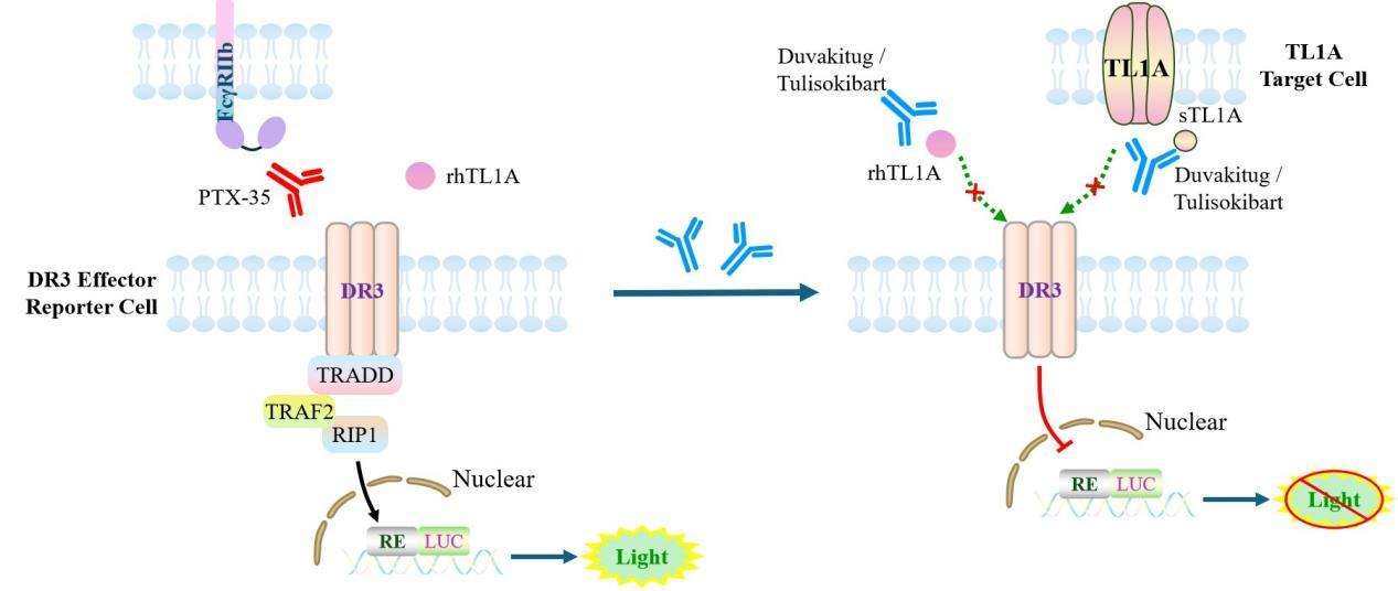
Figure 1. Schematic Diagram of the DR3 Effector Reporter Cell Model
| Classification | Co-Stimulatory |
| Family | Tumor necrosis factor superfamily |
| Gene Name | TNFRSF25 |
| Gene Aliases | TNFRSF12;DR3;TRAMP;WSL-1;LARD;WSL-LR;DDR3;TR3;APO-3 |
| Gene ID | 8718 |
| Accession Number | NM_003790.3 |
| UniProt Number | Q93038 |
| Protein Name | Tumor necrosis factor receptor superfamily member 25 |
| Protein Aliases | Apo-3;Apoptosis-inducing receptor AIR;Apoptosis-mediating receptor DR3;Apoptosis-mediating receptor TRAMP;Death receptor 3 |
| Target Species | Human |
| Host cell | Jurkat E6.1 |
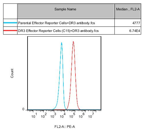
Figure 2. Recombinant DR3 Effector Reporter Cell stably expressing DR3.
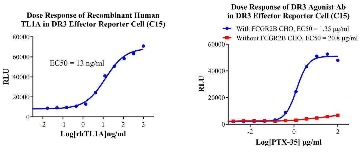
Figure 3. Dose Response of Recombinant Human TL1A in DR3 Effector Reporter Cell(C15). Dose Response of DR3 Agonist Ab in DR3 Effector Reporter Cell(C15)
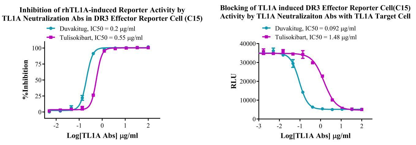
Figure 4. Inhibition of rhTL1A-induced Reporter Activity by TL1A Neutralization Abs in DR3 Effector Reporter Cell(C15). Blocking of hTL1A induced DR3 Effector Reporter Cell(C15) Activity by TL1A Neutralizaiton Abs with TL1A Target Cell.
Cell Passage Procedures
1.This cell line grows in suspension.
2.Upon receipt, cells should be thawed immediately or stored in liquid nitrogen until use.
3.Before thawing, pre-warm the water bath and culture medium to 37 °C, and prepare a small amount of dry ice.
4.Remove the cryovial from storage and transport it to the cell culture laboratory on dry ice.
5.Rapidly thaw the cells in a 37 °C water bath. Once the cells are completely thawed, spray the cryovial with 70% ethanol for disinfection and transfer it to a biosafety cabinet.
6.Add 10 mL of pre-warmed culture medium into a 15 mL centrifuge tube. Transfer the contents of the cryovial into the tube and centrifuge at 1000 rpm for 5 minutes.
7.Carefully discard the supernatant. Resuspend the cell pellet in 5 mL of pre-warmed culture medium by gentle pipetting. Immediately perform cell counting and adjust the cell density to 3–6 × 10⁵ cells/mL based on the counting results, then transfer the cells into a culture flask.
8.Count the cells every 1–2 days. When the cell density exceeds 1 × 10⁶ cells/mL, passage the cells promptly or add fresh culture medium. Maintain the cell density between 2 × 10⁵ and 1 × 10⁶ cells/mL.
Suspension Cell Cryopreservation Procedure:
1.Collect 8 × 10⁶ cells, centrifuge, and discard the supernatant.
2.Add 1 mL of cell freezing medium (90% FBS + 10% DMSO) and gently pipette to mix thoroughly. Transfer the suspension into a cryovial.
3.Immediately place the cryovial into a controlled-rate freezing container (Nalgene 5100-0001), fill with isopropanol up to the indicated level, and store at −80 °C.
4.After 24 hours, transfer the cryovial to liquid nitrogen for long-term storage.
Related products
CHO-K1 Human CCR4 Cell Line
HEK293 Human NK1R CRE-Luc Cell Line
Raji-Luc-GFP
Jurkat E6.1-Luc
THP-1-GFP
THP-1-Luc
Raji-GFP
Raji-Luc
Jurkat E6.1-GFP
HEK293 Human GAL4-Luc Cell
We Are Pleased to Announce: Global Commercial Licensing Rights for Jurkat E6.1, CHO-K1, and HEK293 Cell Lines Officially Secured.
Explore