

Jurkat E6.1 Human PD1&41BB Dual Effector Reporter Cell
Cat. No: RQP74172
Size: 1 vial of frozen cells (>1E6 per vial in 1 mL)
Unit Price: Contact For Pricing
Product Info
Description
Biological Information
Assay Data
Cell Culture
| Cat. No | RQP74172 |
| Product Name | Jurkat E6.1 Human PD1&41BB Dual Effector Reporter Cell |
| Product Type | Reporter Cell |
| Culture Properties | suspension |
| Stability | 32passages (in-house test, that not means the cell line will be instable beyond the passages we tested.) |
| Mycoplasma Status | Negative |
| Culture Medium | RPMI-1640+10%FBS+1μg/ml puromycin+800μg/ml Hygromycin B+10μg/ml blasticidin |
| Freeze Medium | 90% FBS+10% DMSO |
| Storage Conditions | Liquid nitrogen immediately upon delivery |
| Application | Functional(Report Gene) Assay |
For research use only. Not intended for human or animal clinical trials, therapeutic or diagnostic use.
Tumor cells can evade recognition and killing by the host immune system through immune checkpoint receptors. Therefore, blocking these immune checkpoint receptors may represent a broadly effective tumor immunotherapy approach. Currently, although anti-PD-1/PD-L1 antibodies are relatively mature, similar to anti-CTLA4 antibodies, patient overall response rates remain low due to the presence of resistance, making the search for new tumor immunotherapy targets urgent.
Programmed Cell Death Protein 1 (PD-1), a receptor expressed on activated T cells, binds to its ligands PD-L1 and PD-L2 to negatively regulate immune responses. PD-1 ligands are present in most cancers, and the PD-1:PD-L1/2 interaction inhibits T cell activity, allowing cancer cells to escape immune surveillance. The PD-1/PD-L1 signaling pathway is an important component of tumor immune suppression, capable of inhibiting T lymphocyte activation, enhancing tumor cell immune tolerance, and thus enabling tumor immune escape. Binding of PD-1 to PD-L1 weakens T cell-mediated immune surveillance, leading to impaired immune responses and even T cell apoptosis. PD-1/PD-L1 inhibitors can relieve immune suppression of anti-tumor T cells, resulting in T cell proliferation, infiltration into the tumor microenvironment, and induction of anti-tumor responses. The PD-1:PD-L1/2 pathway also participates in regulating autoimmune responses, making these proteins promising therapeutic targets for various cancers as well as multiple sclerosis, arthritis, lupus, and type 1 diabetes.
4-1BB [also known as CD137 or TNF receptor superfamily member 9 (TNFRSF9)] is a glycosylated type I membrane protein belonging to the TNFR superfamily, composed of four extracellular cysteine-rich pseudo-repetitive domains. The cytoplasmic region of 4-1BB contains TNF receptor-associated factor (TRAF) binding motifs. 4-1BB expression is primarily found on the surface of activated cytotoxic CD8+ T cells and helper CD4+ T cells. Upon activation, 4-1BB expression can be induced in NK cells, B cells, monocytes, and dendritic cells. As an inducible co-stimulatory molecule, 4-1BB enhances T cell responses to antigens. When upregulated and trimerized on T cells by 4-1BBL, 4-1BB recruits TRAF adaptor proteins to the cytoplasmic TRAF binding motif, thereby initiating co-stimulatory signaling. The co-stimulatory activity of 4-1BB has the potential to complement and enhance the anti-tumor activity of PD-1/PD-L1 blockade by improving the function and survival of long-term stimulated CD8+ T cells in the tumor microenvironment and expanding T cell clonality and overall prevalence. Tumor antigen-specific T cells express both PD-1 and 4-1BB, providing further theoretical basis for co-targeting these pathways to amplify tumor-specific T cell responses. Bispecific antibody (bsAb) technology offers the opportunity to generate immunotherapeutic agents with superior or novel properties compared to individual mAbs or their mixtures.
The PD1 4-1BB Dual Effector Reporter Cell reporter gene drug target model effectively simulates the signal transduction process of PD1 and 4-1BB in vivo, as illustrated in the figure below.
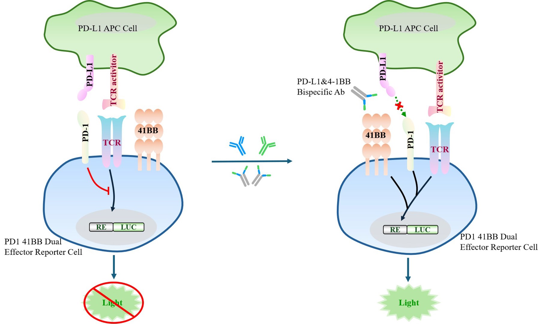
Figure 1. Schematic diagram of the PD1 4-1BB Dual Effector Reporter Cell model.
| Classification | Combination |
| Family | CD28 family |
| Gene Name | PDCD1 |
| Gene Aliases | CD279;PD1;hSLE1;PD-1 |
| Gene ID | 5133 |
| Accession Number | NM_005018.3 |
| UniProt Number | Q15116 |
| Protein Name | Protein PD-1;hPD-1 |
| Protein Aliases | N/A |
| Family-2 | Tumor necrosis factor receptor superfamily |
| Gene Name-2 | 4-1BB |
| Gene Aliases-2 | CD137;TNFRSF9;ILA |
| Gene ID-2 | 3604 |
| Accession Number-2 | NM_001561.6 |
| UniProt Number-2 | Q07011 |
| Protein Name-2 | Tumor necrosis factor receptor superfamily member 9 |
| Protein Aliases-2 | 4-1BB ligand receptor;CDw137;T-cell antigen 4-1BB homolog;T-cell antigen ILA |
| Target Species | Human |
| Host cell | Jurkat E6.1 |
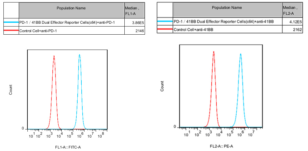
Figure 2. Recombinant PD1 41BB Dual Effector Reporter Cell stably expressing PD-1 and 41BB.
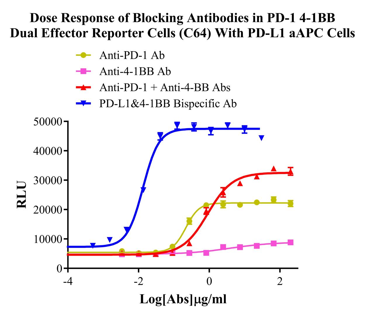
Figure 3. Dose Response of Blocking Antibodies in PD-1 4-1BB Dual Effector Reporter Cells (C64) With PD-L1 aAPC Cells.
Cell Passage Procedures
1.This cell line grows in suspension.
2.Upon receipt, cells should be thawed immediately or stored in liquid nitrogen until use.
3.Before thawing, pre-warm the water bath and culture medium to 37 °C, and prepare a small amount of dry ice.
4.Remove the cryovial from storage and transport it to the cell culture laboratory on dry ice.
5.Rapidly thaw the cells in a 37 °C water bath. Once the cells are completely thawed, spray the cryovial with 70% ethanol for disinfection and transfer it to a biosafety cabinet.
6.Add 10 mL of pre-warmed culture medium into a 15 mL centrifuge tube. Transfer the contents of the cryovial into the tube and centrifuge at 1000 rpm for 5 minutes.
7.Carefully discard the supernatant. Resuspend the cell pellet in 5 mL of pre-warmed culture medium by gentle pipetting. Immediately perform cell counting and adjust the cell density to 3–6 × 10⁵ cells/mL based on the counting results, then transfer the cells into a culture flask.
8.Count the cells every 1–2 days. When the cell density exceeds 1 × 10⁶ cells/mL, passage the cells promptly or add fresh culture medium. Maintain the cell density between 2 × 10⁵ and 1 × 10⁶ cells/mL.
Suspension Cell Cryopreservation Procedure:
1.Collect 8 × 10⁶ cells, centrifuge, and discard the supernatant.
2.Add 1 mL of cell freezing medium (90% FBS + 10% DMSO) and gently pipette to mix thoroughly. Transfer the suspension into a cryovial.
3.Immediately place the cryovial into a controlled-rate freezing container (Nalgene 5100-0001), fill with isopropanol up to the indicated level, and store at −80 °C.
4.After 24 hours, transfer the cryovial to liquid nitrogen for long-term storage.
Related products
CHO-K1 Human CCR4 Cell Line
HEK293 Human NK1R CRE-Luc Cell Line
Raji-Luc-GFP
Jurkat E6.1-Luc
THP-1-GFP
THP-1-Luc
Raji-GFP
Raji-Luc
Jurkat E6.1-GFP
HEK293 Human GAL4-Luc Cell
We Are Pleased to Announce: Global Commercial Licensing Rights for Jurkat E6.1, CHO-K1, and HEK293 Cell Lines Officially Secured.
Explore