

HEK293 Human OSMR Effector Reporter Cell
Cat. No: RQP74157
Size: 1 vial of frozen cells (>1E6 per vial in 1 mL)
Unit Price: Contact For Pricing
Product Info
Description
Biological Information
Assay Data
Cell Culture
| Cat. No | RQP74157 |
| Product Name | HEK293 Human OSMR Effector Reporter Cell |
| Product Type | Reporter Cell |
| Culture Properties | Adherent |
| Stability | 32passages (in-house test, that not means the cell line will be instable beyond the passages we tested.) |
| Mycoplasma Status | Negative |
| Culture Medium | DMEM+10%FBS+200μg/ml Hygromycin B+2μg/ml puromycin+5μg/ml blasticidin |
| Freeze Medium | 90% FBS+10% DMSO |
| Storage Conditions | Liquid nitrogen immediately upon delivery |
| Application | Functional(Report Gene) Assay |
For research use only. Not intended for human or animal clinical trials, therapeutic or diagnostic use.
Oncostatin M (OSM) is a pleiotropic cytokine belonging to the Interleukin-6 (IL-6) family; it participates in various inflammatory processes, such as wound healing, liver regeneration, and bone remodeling. In addition to OSM, members of the IL-6 family include IL-6, Leukemia Inhibitory Factor (LIF), IL-11, IL-27, IL-31, Cardiotrophin-1 (CT-1), Ciliary Neurotrophic Factor (CNTF), and Cardiotrophin-like Cytokine 1 (CLCF1). OSM is widely expressed *in vivo*, and it can be produced by a variety of immune cells, including T cells, monocytes/macrophages, and neutrophils.
The receptor complexes of the IL-6 receptor family all contain the GP130 subunit. OSM receptor complexes are heterodimers; based on the identity of the second subunit within the complex, they can be classified into two types: Type I and Type II. The Type I OSM receptor complex consists of the α-subunit GP130 and the β-subunit LIFRβ (LIF Receptor β-subunit), whereas the Type II OSM receptor complex consists of the α-subunit GP130 and the β-subunit OSMRβ (OSM Receptor β-subunit). When OSM and the two subunits of the OSM receptor complex are simultaneously present, OSM first forms a low-affinity heterodimer with GP130; this heterodimer subsequently recruits and binds to either OSMR or LIFR, thereby activating various signaling pathways, including the JAK/STAT, MAPK, JNK, and PI3K/AKT pathways.
The HEK293 Human OSMR Effector Reporter Cell Model—effectively simulates the signal transduction process of OSMR *in vivo*. The underlying principle is illustrated in the figure below.
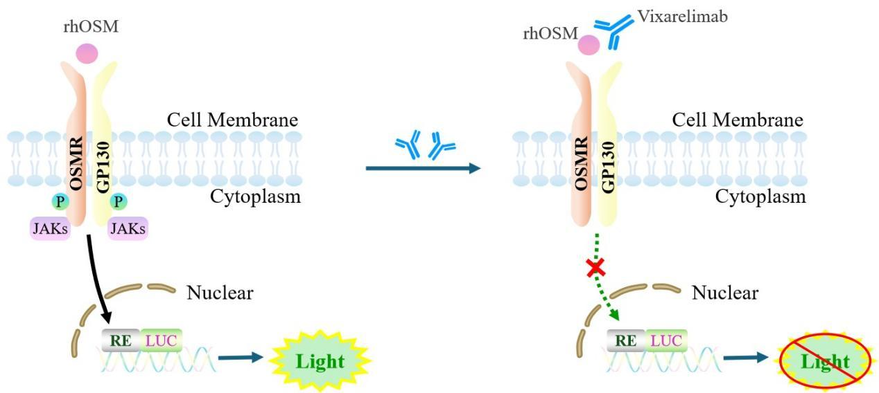
Figure 1. Schematic Diagram of the HEK293 Human OSMR Effector Reporter Cell Model
| Classification | Cytokine&Growth Factor |
| Family | type I cytokine receptor family. Type 2 subfamily |
| Gene Name | OSMR |
| Gene Aliases | OSMRB;OSMRbeta |
| Gene ID | 9180 |
| Accession Number | NM_003999.3 |
| UniProt Number | Q99650 |
| Protein Name | IL-31 receptor subunit beta; IL-31R subunit beta; IL-31R-beta; IL-31RB |
| Protein Aliases | |
| Target Species | Human |
| Host cell | HEK293 |
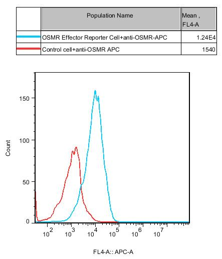
Figure 2. Recombinant OSMR Effector Reporter Cell stably expressing OSMR.
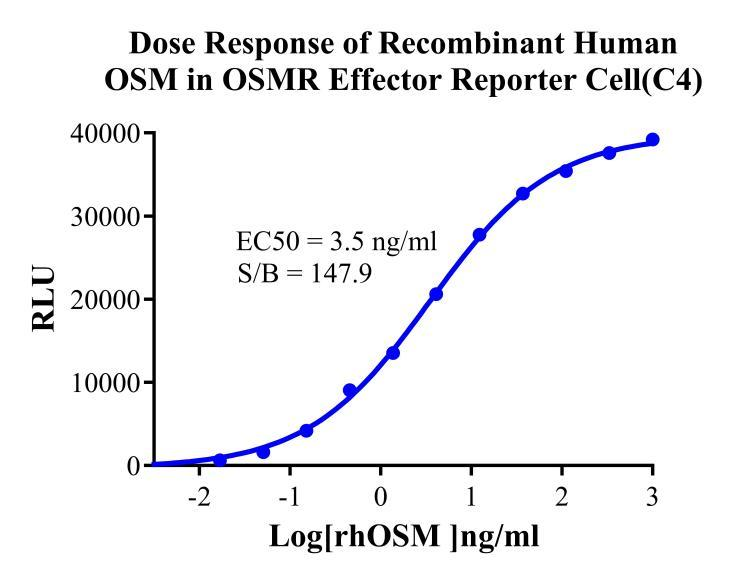
Figure 3.Dose Response of Recombinant Human OSM in OSMR Effector Reporter Cell(C4).
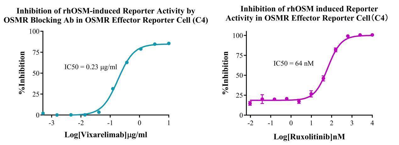
Figure 4. Inhibition of rhOSM-induced Reporter Activity by OSMR Blocking Ab in OSMR Effector Reporter Cell (C4). Inhibition of rhOSM induced Reporter Activity in OSMR Effector Reporter Cell(C4).
Cell Resuscitation
1)Rapidly thaw the frozen cells in a 37 °C water bath for approximately 60 seconds. Once thawed (which may take slightly less or more than 60 seconds), immediately transfer the cell suspension from the cryovial into a 15 mL centrifuge tube containing 10 mL of pre-warmed HEK293 Human OSMR Effector Reporter Cell complete culture medium.
2)Centrifuge cells at 1000 rpm for 5 min to remove medium, then resuspend cells in 5 mL of pre-warmed complete medium.
3)Transfer the cell suspension into a T25 culture flask and incubate at 37 °C with 5% CO₂.
4)After approximately 24–36 hours, replace the medium or passage the cells to remove non-adherent dead cells.
Subculturing procedure
1)When the cell density reaches the appropriate confluency for passaging, wash the cells with PBS, then add 1 mL trypsin to detach the cells. When more than 80% of the cells detach upon gently tapping the culture flask, add complete culture medium to terminate digestion. Gently pipette to obtain a single-cell suspension, transfer to a 15 mL centrifuge tube, and centrifuge at 1000 rpm for 5 minutes.
2)Discard supernatant after centrifugation. Resuspend cells in fresh medium to a single-cell suspension and transfer to a new culture flask for continued growth.
Cell Freezing
After trypsinization and centrifugation of cells from each T75 flask or 10 cm culture dish, discard the supernatant. Add 2 mL of cryopreservation medium (90% FBS + 10% DMSO), gently resuspend thoroughly, and aliquot into two cryovials. Immediately place the cryovials into a controlled-rate freezing container (e.g., Nalgene 5100-0001), fill with isopropanol to the indicated level, and store at −80 °C. After 24 hours, transfer the cryovials to liquid nitrogen for long-term storage.
Related products
CHO-K1 Human CCR4 Cell Line
HEK293 Human NK1R CRE-Luc Cell Line
Raji-Luc-GFP
Jurkat E6.1-Luc
THP-1-GFP
THP-1-Luc
Raji-GFP
Raji-Luc
Jurkat E6.1-GFP
HEK293 Human GAL4-Luc Cell
We Are Pleased to Announce: Global Commercial Licensing Rights for Jurkat E6.1, CHO-K1, and HEK293 Cell Lines Officially Secured.
Explore