

HEK293 Human NOD2 Effector Reporter Cell
Cat. No: RQP74536
Size: 1 vial of frozen cells (>1E6 per vial in 1 mL)
Unit Price: Contact For Pricing
Product Info
Description
Biological Information
Assay Data
Cell Culture
| Cat. No | RQP74536 |
| Product Name | HEK293 Human NOD2 Effector Reporter Cell |
| Product Type | Reporter Cell |
| Culture Properties | Adherent |
| Stability | 32passages (in-house test, that not means the cell line will be instable beyond the passages we tested.) |
| Mycoplasma Status | Negative |
| Culture Medium | MEM +10%FBS + 1% NEAA+ 1mM NaP+1 μg/ml Puromycin+ 100 μg/ml Hygromycin B |
| Freeze Medium | 90% FBS+10% DMSO |
| Storage Conditions | Liquid nitrogen immediately upon delivery |
| Application | Functional(Report Gene) Assay |
For research use only. Not intended for human or animal clinical trials, therapeutic or diagnostic use.
NOD-like receptors (NLRs) constitute a critical class of intracellular pattern recognition receptors within the innate immune system; their core function is to sense pathogen-associated molecular patterns (PAMPs) or danger-associated molecular patterns (DAMPs), thereby initiating an immune response. Based on their distinct N-terminal domains, the human NLR family can be broadly categorized into four major subfamilies: NLRA (e.g., CIITA), NLRB (e.g., NAIP), NLRC (e.g., NOD1, NOD2, NLRC3/4/5), and NLRP (containing a PYD domain; e.g., NLRP1, NLRP3). Among these, NOD1 and NOD2 both belong to the NLRC subfamily; they primarily recognize fragments of bacterial cell wall peptidoglycans and serve as classic sensors in antibacterial innate immunity.
Compared to NOD1, the most distinctive structural feature of NOD2 (full name: Nucleotide-binding oligomerization domain-containing protein 2) is the presence of two tandem CARD domains at its N-terminus (whereas NOD1 possesses only one CARD domain); this structural difference dictates their distinct ligand recognition specificities and signaling intensities. NOD2 is predominantly expressed in monocytes/macrophages, dendritic cells, Paneth cells, and intestinal epithelial cells, among others. NOD2 specifically recognizes muramyl dipeptide (MDP)—a component of bacterial cell wall peptidoglycans—which represents the minimal immunoactive structural unit shared by nearly all Gram-positive and Gram-negative bacteria. Consequently, the ligand repertoire of NOD2 is broader than that of NOD1. Loss-of-function mutations in the *NOD2* gene (particularly L1007fs, G908R, and R702W) constitute the strongest genetic risk factors for Crohn's disease, underscoring the critical role of NOD2 in maintaining intestinal immune tolerance and barrier function. Conversely, gain-of-function mutations (e.g., E383K) are associated with Blau syndrome—a condition characterized by early-onset granulomatous arthritis, uveitis, and dermatitis—thereby illustrating the spectrum of diseases resulting from dysregulated NOD2 signaling, ranging from immunodeficiency to excessive inflammation.
The HEK293 Human NOD2 Effector Reporter Cell model effectively mimics the in vivo MET signaling transduction process. The principle is illustrated in the figure below.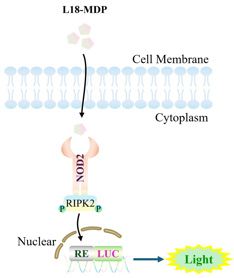
Figure 1. Schematic diagram of the HEK293 Human NOD2 Effector Reporter Cell model
| Classification | TLR |
| Family | NOD1-NOD2 family |
| Gene Name | NOD2 |
| Gene Aliases | IBD1;CARD15;BLAU;CD;PSORAS1;CLR16.3;NLRC2 |
| Gene ID | 64127 |
| Accession Number | NM_001370466.1 |
| UniProt Number | Q9HC29 |
| Protein Name | Nucleotide-binding oligomerization domain-containing protein 2 |
| Protein Aliases | Caspase recruitment domain-containing protein 15;Inflammatory bowel disease protein 1 |
| Target Species | Human |
| Host cell | HEK293 |
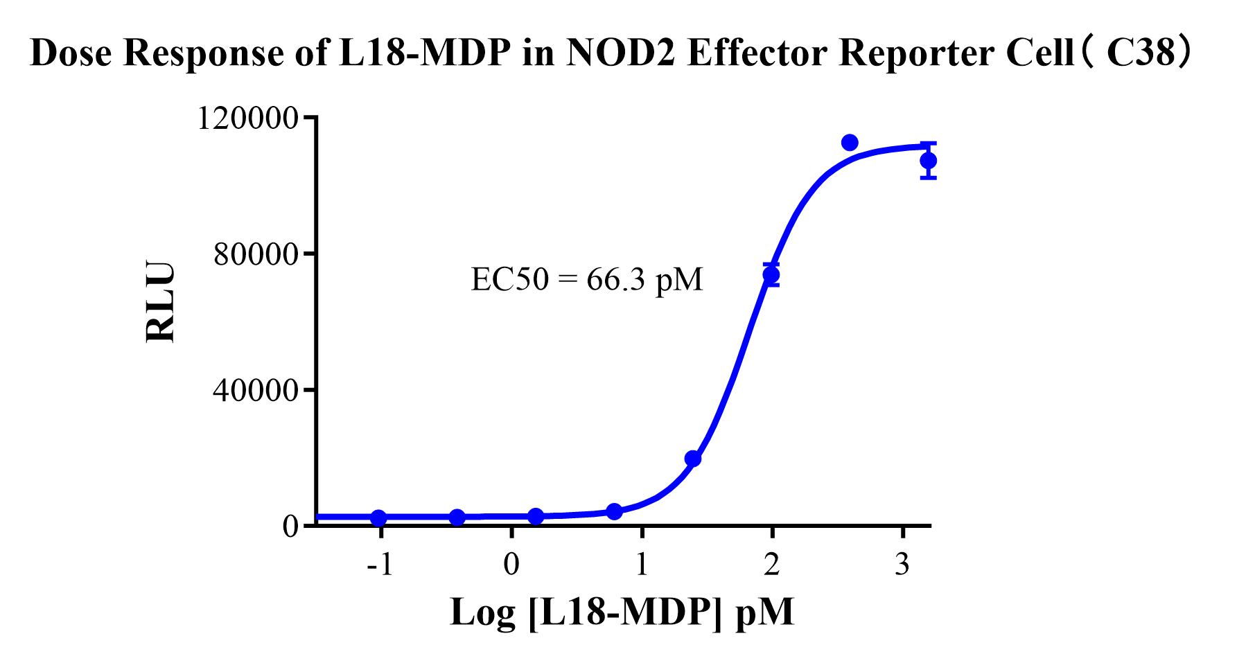
Figure 2. Dose Response of L18-MDP in NOD2 Effector Reporter Cell(C38).
Cell Resuscitation
1)Rapidly thaw the frozen cells in a 37 °C water bath for approximately 60 seconds. Once thawed (which may take slightly less or more than 60 seconds), immediately transfer the cell suspension from the cryovial into a 15 mL centrifuge tube containing 10 mL of pre-warmed HEK293 Human NOD2 Effector Reporter Cell complete culture medium.
2)Centrifuge cells at 1000 rpm for 5 min to remove medium, then resuspend cells in 5 mL of pre-warmed complete medium.
3)Transfer the cell suspension into a T25 culture flask and incubate at 37 °C with 5% CO₂.
4)After approximately 24–36 hours, replace the medium or passage the cells to remove non-adherent dead cells.
Subculturing procedure
1)When the cell density reaches the appropriate confluency for passaging, wash the cells with PBS, then add 1 mL trypsin to detach the cells. When more than 80% of the cells detach upon gently tapping the culture flask, add complete culture medium to terminate digestion. Gently pipette to obtain a single-cell suspension, transfer to a 15 mL centrifuge tube, and centrifuge at 1000 rpm for 5 minutes.
2)Discard supernatant after centrifugation. Resuspend cells in fresh medium to a single-cell suspension and transfer to a new culture flask for continued growth.
Cell Freezing
After trypsinization and centrifugation of cells from each T75 flask or 10 cm culture dish, discard the supernatant. Add 2 mL of cryopreservation medium (90% FBS + 10% DMSO), gently resuspend thoroughly, and aliquot into two cryovials. Immediately place the cryovials into a controlled-rate freezing container (e.g., Nalgene 5100-0001), fill with isopropanol to the indicated level, and store at −80 °C. After 24 hours, transfer the cryovials to liquid nitrogen for long-term storage.
Related products
CHO-K1 Human CCR4 Cell Line
HEK293 Human NK1R CRE-Luc Cell Line
Raji-Luc-GFP
Jurkat E6.1-Luc
THP-1-GFP
THP-1-Luc
Raji-GFP
Raji-Luc
Jurkat E6.1-GFP
HEK293 Human GAL4-Luc Cell
We Are Pleased to Announce: Global Commercial Licensing Rights for Jurkat E6.1, CHO-K1, and HEK293 Cell Lines Officially Secured.
Explore