

CHO-K1 Human OMSR&IL6ST Dimerization Cell
Cat. No: RQP74464
Size: 1 vial of frozen cells (>1E6 per vial in 1 mL)
Unit Price: Contact For Pricing
Product Info
Description
Biological Information
Assay Data
Cell Culture
| Cat. No | RQP74464 |
| Product Name | CHO-K1 Human OMSR&IL6ST Dimerization Cell |
| Product Type | Reporter Cell |
| Culture Properties | Adherent |
| Stability | 32passages (in-house test, that not means the cell line will be instable beyond the passages we tested.) |
| Mycoplasma Status | Negative |
| Culture Medium | F12K+10%FBS+ 5 μg/ml Puromycin+5 μg/ml Blasticidin |
| Freeze Medium | 90% FBS+10% DMSO |
| Storage Conditions | Liquid nitrogen immediately upon delivery |
| Application | Functional(Report Gene) Assay |
For research use only. Not intended for human or animal clinical trials, therapeutic or diagnostic use.
Oncostatin M (OSM) is a pleiotropic cytokine belonging to the Interleukin-6 (IL-6) family; it participates in various inflammatory responses, such as wound healing, liver regeneration, and bone remodeling. In addition to OSM, members of the IL-6 family include IL-6, Leukemia Inhibitory Factor (LIF), IL-11, IL-27, IL-31, Cardiotrophin-1 (CT-1), Ciliary Neurotrophic Factor (CNTF), and Cardiotrophin-like Cytokine 1 (CLCF1). OSM is widely expressed *in vivo*, and it can be produced by a variety of immune cells, including T cells, monocytes/macrophages, and neutrophils.
The receptor complexes of the IL-6 receptor family all contain the GP130 subunit. The receptor complexes for OSM are heterodimers; based on the identity of the second subunit within the complex, these receptors can be classified into two types: Type I and Type II. The Type I OSM receptor complex consists of the α-subunit GP130 and the β-subunit LIFRβ (LIF Receptor β-subunit), whereas the Type II OSM receptor complex consists of the α-subunit GP130 and the β-subunit OSMRβ (OSM Receptor β-subunit). When OSM and the two subunits of its receptor complex are simultaneously present, OSM first forms a low-affinity heterodimer with GP130; this heterodimer then recruits and binds to either OSMR or LIFR, thereby activating multiple signaling pathways, including the JAK/STAT, MAPK, JNK, and PI3K/AKT pathways.
The OSMR & IL6ST Dimerization CHO Reporter Gene Drug Target Model effectively simulates the *in vivo* signal transduction process of OSM; the underlying principle is illustrated in the figure below.
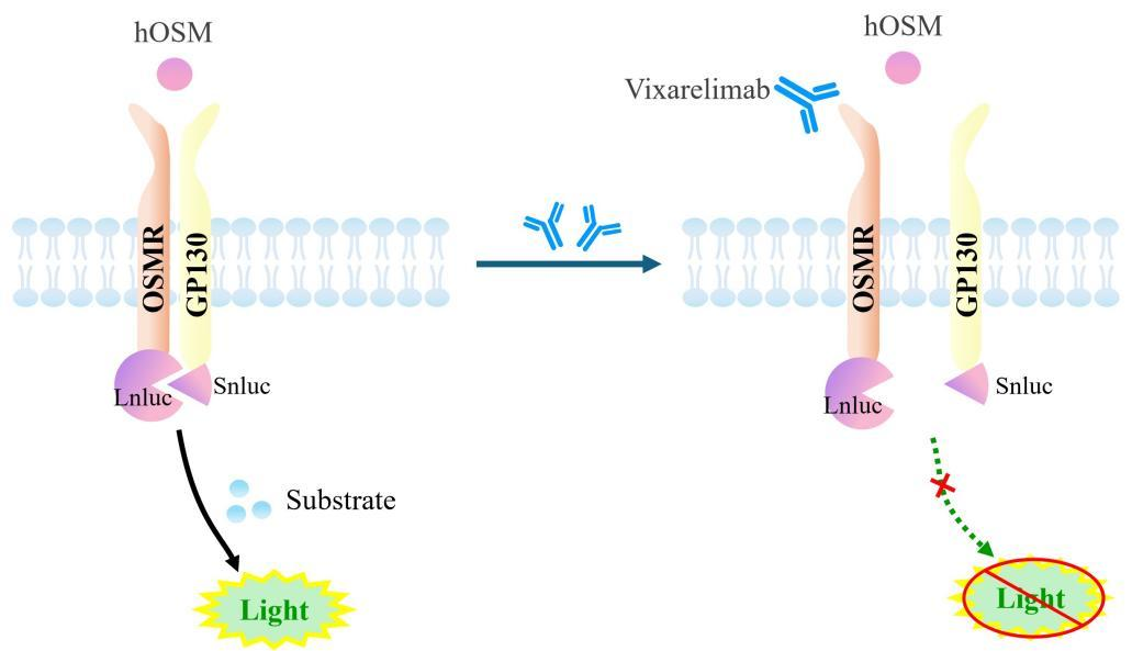
Figure 1. Schematic Diagram of the OSMR & IL6ST Dimerization CHO Cell Model
| Classification | Cytokine&Growth Factor |
| Family | type I cytokine receptor family. Type 2 subfamily |
| Gene Name | OSMR |
| Gene Aliases | OSMRB;OSMRbeta |
| Gene ID | 9180 |
| Accession Number | NM_003999.3 |
| UniProt Number | Q99650 |
| Protein Name | IL-31 receptor subunit beta; IL-31R subunit beta; IL-31R-beta; IL-31RB |
| Protein Aliases | N/A |
| Family-2 | type I cytokine receptor family. Type 2 subfamily |
| Gene Name-2 | IL6ST |
| Gene Aliases-2 | GP130;CD130;sGP130;IL-6RB |
| Gene ID-2 | 3572 |
| Accession Number-2 | NM_002184.4 |
| UniProt Number-2 | P40189 |
| Protein Name-2 | IL-6 receptor subunit beta; IL-6R subunit beta; IL-6R-beta; IL-6RB |
| Protein Aliases-2 | CDw130;Interleukin-6 signal transducer;gp130 |
| Target Species | Human |
| Host cell | CHO-K1 |
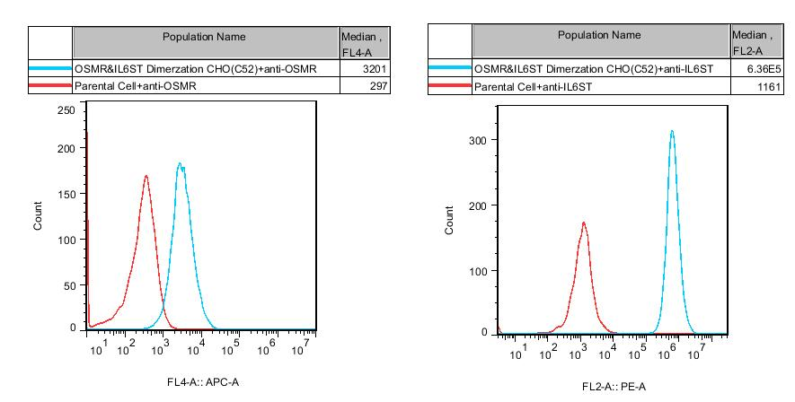
Figure 2. Recombinant OSMR&IL6ST Dimerization CHO stably expressing OSMR&IL6ST.
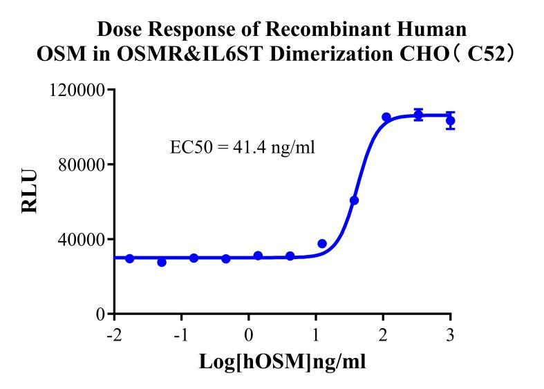
Figure 3. Dose Response of Recombinant Human OSM in OSMR&IL6ST Dimerization CHO( C52).
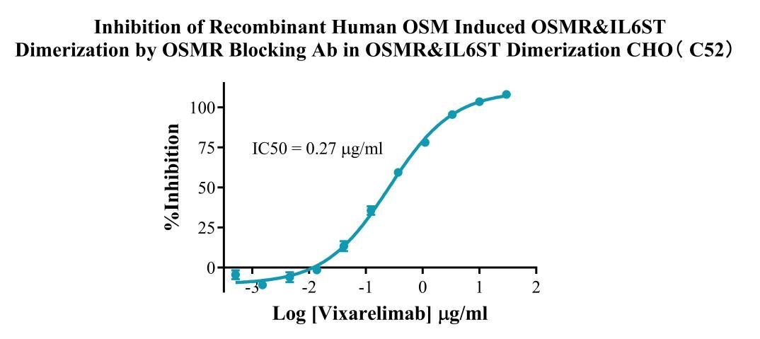
Figure 4. Inhibition of Recombinant Human OSM induced OSMR&IL6ST Dimerization by OSMR Blocking Ab in OSMR&IL6ST Dimerization CHO (C52).
Cell Resuscitation
1)Rapidly thaw the frozen cells in a 37 °C water bath for approximately 60 seconds. Once thawed (which may take slightly less or more than 60 seconds), immediately transfer the cell suspension from the cryovial into a 15 mL centrifuge tube containing 10 mL of pre-warmed CHO-K1 Human OMSR&IL6ST Dimerization Cell complete culture medium.
2)Centrifuge cells at 1000 rpm for 5 min to remove medium, then resuspend cells in 5 mL of pre-warmed complete medium.
3)Transfer the cell suspension into a T25 culture flask and incubate at 37 °C with 5% CO₂.
4)After approximately 24–36 hours, replace the medium or passage the cells to remove non-adherent dead cells.
Subculturing procedure
1)When the cell density reaches the appropriate confluency for passaging, wash the cells with PBS, then add 1 mL trypsin to detach the cells. When more than 80% of the cells detach upon gently tapping the culture flask, add complete culture medium to terminate digestion. Gently pipette to obtain a single-cell suspension, transfer to a 15 mL centrifuge tube, and centrifuge at 1000 rpm for 5 minutes.
2)Discard supernatant after centrifugation. Resuspend cells in fresh medium to a single-cell suspension and transfer to a new culture flask for continued growth.
Cell Freezing
After trypsinization and centrifugation of cells from each T75 flask or 10 cm culture dish, discard the supernatant. Add 2 mL of cryopreservation medium (90% FBS + 10% DMSO), gently resuspend thoroughly, and aliquot into two cryovials. Immediately place the cryovials into a controlled-rate freezing container (e.g., Nalgene 5100-0001), fill with isopropanol to the indicated level, and store at −80 °C. After 24 hours, transfer the cryovials to liquid nitrogen for long-term storage.
Related products
CHO-K1 Human CCR4 Cell Line
HEK293 Human NK1R CRE-Luc Cell Line
Raji-Luc-GFP
Jurkat E6.1-Luc
THP-1-GFP
THP-1-Luc
Raji-GFP
Raji-Luc
Jurkat E6.1-GFP
HEK293 Human GAL4-Luc Cell
We Are Pleased to Announce: Global Commercial Licensing Rights for Jurkat E6.1, CHO-K1, and HEK293 Cell Lines Officially Secured.
Explore